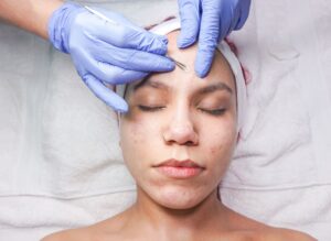
Histology of acne scars guides treatment planning because different scar types have distinct structural characteristics that determine which procedures will actually work. A dermatologist examining acne scars under magnification sees fundamentally different damage patterns—some scars are narrow V-shaped depressions extending deep into the dermis, others are broad undulating surfaces, and still others are box-like indentations with vertical walls. These microscopic differences aren’t just academic; they directly determine whether a patient will respond to laser treatment, microneedling, injectable fillers, or surgical techniques.
For example, a deep ice-pick scar that penetrates through the entire dermis thickness won’t respond to superficial microneedling the way a rolling scar will, because the histological architecture is completely different. Understanding acne scar histology shifts treatment from guesswork to precision medicine. This article explains how dermatologists use histological classification to select treatments, what the major scar types look like under examination, how laboratory findings influence therapy selection, and why a one-size-fits-all approach to acne scarring almost always disappoints patients. We’ll cover the three primary atrophic scar categories, the histopathological findings that predict treatment success, evidence-based protocols for each scar morphology, and emerging multi-modal strategies that combine early intervention with targeted procedures.
Table of Contents
- What Does Histology Reveal About Acne Scar Structure?
- The Three Major Types of Atrophic Acne Scars and Their Histological Characteristics
- How Histopathological Findings Predict Treatment Outcomes
- Treatment Planning by Scar Morphology: From Diagnosis to Procedure Selection
- Emerging Morphology-Driven Algorithms and Combination Therapy
- Multi-Modal Treatment Integration and Practical Application
- The Future of Histology-Guided Acne Scar Treatment
- Conclusion
What Does Histology Reveal About Acne Scar Structure?
Histopathological evaluations of acne scars measure several key parameters that dermatologists use to decide on treatment: fibroblast activity levels, epidermal thickness variations, and collagen composition and density in the dermis. When a dermatologist takes a tissue sample from scarred skin, laboratory analysis reveals whether the scar involves primarily a loss of collagen (atrophic scar), an excess of collagen (hypertrophic or keloid), or a combination of both. These findings matter because treating a scar with inadequate collagen requires stimulation (lasers, microneedling, radiofrequency), while treating excessive collagen deposition requires different approaches (steroid injections, laser ablation). The histological picture essentially tells the clinician what biological problem needs solving. The structure beneath the surface determines everything about how a scar responds to treatment.
A scar that appears depressed on the skin surface might involve thinned epidermis, loss of dermal collagen, or both. Some scars show fibroblasts that are relatively inactive—these scars have essentially “matured” into stable fibrosis and won’t remodel easily. Other scars show ongoing inflammation or abnormal fibroblast behavior that suggests they might still respond to remodeling therapy. In practical terms, a patient with a 10-year-old ice-pick scar involving mature, stable fibrosis will require more aggressive treatment (potentially surgical excision) than a patient with recent rolling scars still showing signs of active fibroblast dysregulation. The histology tells the story of what happened and what’s most likely to help.
The Three Major Types of Atrophic Acne Scars and Their Histological Characteristics
Ice-pick scars represent 60-70% of atrophic acne scarring and are the most recognizable and most challenging type. Histologically, they appear as V-shaped depressions narrower than 2mm that penetrate through the entire thickness of the dermis, sometimes reaching into subcutaneous tissue. Because of their depth and narrow width, ice-pick scars create a piercing appearance on the skin surface—they look like small puncture wounds. The narrow channel means there’s minimal surrounding dermis to remodel or fill, which is why these scars are historically the most resistant to conventional treatments. Treatment planning for ice-pick scars typically involves accepting that many conventional procedures won’t work well and reserving punch techniques or surgical approaches as last resorts. Rolling scars comprise 15-25% of atrophic scars and have distinctly different histology. These scars are wider (4-6mm diameter), with an undulating or wavelike appearance caused by fibrous anchoring that tethers the dermis to underlying subcutaneous tissue.
Histologically, rolling scars show broader areas of collagen loss with abnormal collagen organization, and the tethered appearance reflects scar tissue that’s literally pulling the skin down. The key difference from ice-pick scars: rolling scars involve a larger surface area of abnormal tissue and respond well to subcision (cutting the fibrous tethers beneath the skin), filler injections, and microneedling because there’s more dermal surface available for remodeling. A patient with rolling scars is typically a good candidate for combination therapy, whereas ice-pick scar patients often face limited options. Boxcar scars represent 20-30% of atrophic scars and occupy a middle ground histologically. They appear as round or polygonal depressions with steep vertical walls, a flat base, and dimensions of 1-4mm in diameter with 0.1-0.5mm depth. Unlike rolling scars with their undulating appearance, boxcar scars look more like small pits or craters with clearly defined borders. Histologically, boxcar scars involve collagen loss in a more circumscribed area with defined margins, rather than the broad tethering seen in rolling scars or the deep channels of ice-pick scars. However, if a boxcar scar is deep enough to have vertical walls, it may require surgical techniques (like punch excision) to improve significantly, because the histological barrier of thick fibrous tissue at the base makes non-surgical remodeling less effective.
How Histopathological Findings Predict Treatment Outcomes
Histopathological evaluation identifies specific markers that correlate with treatment success or failure. Dermatologists look for increased fibroblast activity—active, proliferating fibroblasts suggest the scar tissue is still remodeling and might respond to stimulation therapies like lasers or microneedling. Conversely, mature scars with established fibrosis and low fibroblast activity typically require more aggressive intervention because the tissue has “settled” into a stable state and won’t easily change shape. Additionally, histological analysis reveals collagen density and the organization of collagen fibers; disorganized collagen architecture with lower density is more amenable to remodeling and filling strategies than densely packed, well-organized scar tissue.
CO₂ and Er:YAG laser treatments achieve 90% efficacy rates according to histological studies measuring specific outcomes: increased dermal collagen density, extended elastic fiber length, and upregulation of matrix metalloproteinase-3 (MMP-3) gene expression. These laser therapies work by creating controlled dermal injury that triggers the wound-healing cascade, prompting fibroblasts to synthesize new, organized collagen. However—and this is a critical limitation—histological studies showing 90% efficacy typically measure these outcomes in ideal candidates with appropriate scar types. A patient with mature ice-pick scars might see minimal improvement because the laser is trying to stimulate new collagen in a scar type that’s histologically resistant to remodeling. The histology determines whether the patient falls into the group likely to see excellent results or the group where laser therapy will provide only partial improvement.
Treatment Planning by Scar Morphology: From Diagnosis to Procedure Selection
Ice-pick scars demand the most conservative initial approach followed by escalation if needed, precisely because their histology limits options. Since they penetrate deeply through the entire dermis and have minimal surrounding tissue to remodel, non-invasive procedures like microneedling or fractional laser will show limited benefit. The evidence-based approach involves punch techniques as a last resort for deep ice-pick scars resistant to other approaches—meaning the dermatologist essentially removes the scarred channel surgically and lets the skin heal with better collagen organization. Some clinicians combine this with earlier attempts at fractional CO₂ laser or chemical peels, but histological reality dictates that ice-pick scars rarely achieve cosmetically significant improvement without some form of surgical intervention.
Rolling and boxcar scars respond far better to multi-modal approaches because their histology supports remodeling. Microneedling demonstrates the greatest success rate for rolling scars because the procedure creates controlled dermal injury across the broader surface area involved in rolling scars, stimulating organized collagen synthesis. Subcision combined with fillers is the standard approach for rolling scars with significant tethering—cutting the fibrous anchors beneath the skin allows the depressed area to elevate, and injectable fillers provide immediate volume while new collagen organizes underneath. For boxcar scars, the approach depends on depth: superficial boxcar scars (less than 0.5mm deep) may respond well to lasers and microneedling, while deeper boxcar scars may require punch excision similar to ice-pick scars. The histological difference guides the choice: is the scar shallow enough that remodeling and filling can help, or is it deep enough to require surgical redefinition?.
Emerging Morphology-Driven Algorithms and Combination Therapy
Recent research emphasizes that a 2025-2026 emerging strategy applies morphology-driven algorithms that match specific scar types to optimized combinations of procedures. Rather than using a single modality, the protocol selects based on histological classification: CROSS (chemical reconstruction of skin scars) plus laser for ice-pick scars, subcision plus fillers for rolling scars, and tailored laser or punch approaches for boxcar scars. This evolution reflects an important understanding—acne scars aren’t all the same problem, so they shouldn’t all receive the same solution. A limitation of this approach is that it requires dermatologists to accurately identify scar types and understand the evidence for each morphology-specific treatment, which not all clinicians do consistently.
Combined therapy involving early intervention makes a substantial difference. Research from 2025 shows that isotretinoin treatment during active acne combined with early adjunctive procedures like chemical peels and lasers significantly improves outcomes compared to single modality approaches. This means that preventing scarring through aggressive acne management, then intervening early with lasers or other procedures before scars fully mature histologically, produces better results than waiting years to treat established scars. However, if a patient comes in with fully mature scars that have completed remodeling (which typically takes 12-24 months after acne resolves), the window for early intervention has passed and more aggressive procedures become necessary. The histology of mature scars is less responsive simply because the fibroblasts have finished their work and settled into stable fibrosis.

Multi-Modal Treatment Integration and Practical Application
In practice, combining procedures addresses different histological features simultaneously. A patient with mixed rolling and boxcar scarring might benefit from microneedling for the boxcar components while undergoing subcision for the rolling components, potentially followed by filler injection for volume restoration. Some dermatologists perform microneedling followed by topical treatments or fractional laser on the same day to amplify the remodeling response.
Others use combination therapy sequentially—perhaps subcision in one session, then laser resurfacing four weeks later once the skin has healed—to allow each procedure’s biological effects to stabilize before adding the next stimulus. The histological understanding of each scar type allows dermatologists to design patient-specific protocols rather than offering standard, one-size-fits-all approaches. An example of this integration: a patient with predominantly rolling scars and some ice-pick components might start with subcision and filler for the rolling scars (addressing the tethering and volume loss), then proceed to fractional CO₂ laser for overall skin texture improvement, with the understanding that the deeper ice-pick scars will show minimal cosmetic improvement. This approach respects the histological reality that rolling scars and ice-pick scars require different strategies, so trying to use a single procedure to treat both is less effective than acknowledging their differences and designing a multi-modal plan.
The Future of Histology-Guided Acne Scar Treatment
Moving forward, the field is shifting toward more sophisticated assessment techniques—not just visual classification but actual histological or optical coherence tomography (OCT) imaging that reveals subsurface scar architecture before treatment planning. This technology allows dermatologists to see scar depth, collagen density, and tissue organization in detail, moving beyond visual classification into precise anatomical measurement. While histological biopsies have been historically limited to research settings (because taking tissue samples from facial scars creates additional scarring), emerging non-invasive imaging is closing this gap and making histology-guided planning more accessible.
The integration of early intervention during acne treatment, morphology-specific procedures, and emerging diagnostic tools suggests that future acne scar management will be increasingly personalized and precise. Rather than patients cycling through multiple treatments that don’t address their specific scar histology, understanding the underlying structural differences allows clinicians to recommend evidence-based approaches from the start. For patients with acne scarring, this means seeking dermatologists who can accurately classify scars, understand the histological basis of different scar types, and recommend treatment plans specifically designed for their particular scarring pattern rather than generic acne scar protocols.
Conclusion
Histology of acne scars guides treatment planning because the microscopic structure of scar tissue determines how it will respond to different interventions. Ice-pick, rolling, and boxcar scars represent distinct histological patterns—from deep V-shaped channels to undulating tethered surfaces to circumscribed pits—and each requires a different strategic approach.
By understanding what histopathological evaluations reveal about fibroblast activity, collagen composition, and dermal organization, dermatologists can select procedures that actually address the specific structural problem rather than applying generic treatments to all acne scars equally. Moving forward, the combination of accurate scar classification, morphology-specific treatment protocols, and emerging diagnostic imaging will continue to improve outcomes for patients with acne scarring. If you have atrophic acne scars and are considering treatment, seeking a dermatologist who can distinguish between scar types, explain the histological basis of your scarring, and design a multi-modal plan specific to your scar morphology will substantially improve your chances of meaningful improvement.