A biopsy of acne scar tissue is rarely needed for diagnosis—dermatologists can identify scar types through visual examination alone. However, when a biopsy is performed, it reveals crucial microscopic details about the underlying collagen structure and fibroblast activity that determine which treatment approach will be most effective. For example, if a biopsy shows hypertrophic scar tissue with thick, disorganized collagen bundles arranged in whorls, your dermatologist knows that laser therapy or collagen-remodeling treatments will likely be more successful than topical approaches.
This article explores what biopsies reveal about acne scarring at the cellular level, how these findings guide treatment decisions, and which modern therapies are most effective for different scar types. Understanding the histological (microscopic tissue) findings from biopsies helps explain why one treatment works better than another for individual patients. Rather than guessing which approach suits your scars, the cellular evidence provides a roadmap. This article covers when biopsies are actually indicated, what pathologists look for under the microscope, how findings correlate with scar severity, and the latest treatments informed by these cellular insights.
Table of Contents
- Why Are Biopsies Rarely Performed for Routine Acne Scar Diagnosis?
- What Histological Features Do Biopsies Reveal About Scar Structure?
- How Do Scar Types Correlate with Microscopic Findings?
- How Do Biopsy Findings Guide Treatment Selection?
- What Advanced Biopsy Findings Inform Emerging Therapies?
- The Role of Scar Severity Grading in Treatment Planning
- Future Perspectives: How Biopsy Science Shapes Tomorrow’s Scar Treatments
- Conclusion
Why Are Biopsies Rarely Performed for Routine Acne Scar Diagnosis?
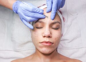
Acne scarring is diagnosed almost entirely through clinical examination—a dermatologist examines your skin visually and gathers information about your acne history. A biopsy is considered only when there is diagnostic uncertainty, such as distinguishing whether a raised scar is a hypertrophic scar (confined to the original injury site) or a keloid scar (extending beyond it). In practice, this situation is uncommon, because experienced dermatologists can differentiate scar types by appearance and palpation alone.
The reason biopsies aren’t routine is straightforward: they are invasive, create a new wound that must heal, take time to process, and add cost without changing the fundamental approach to treatment. Your dermatologist will assess scar depth, type, location, and skin tone to guide therapy selection, all without a biopsy. However, biopsies are valuable in research settings and in complex cases where the clinical picture is unclear—such as when a patient has unusually aggressive scarring that might indicate an underlying collagen disorder or when distinguishing pathological from normal scarring.
What Histological Features Do Biopsies Reveal About Scar Structure?
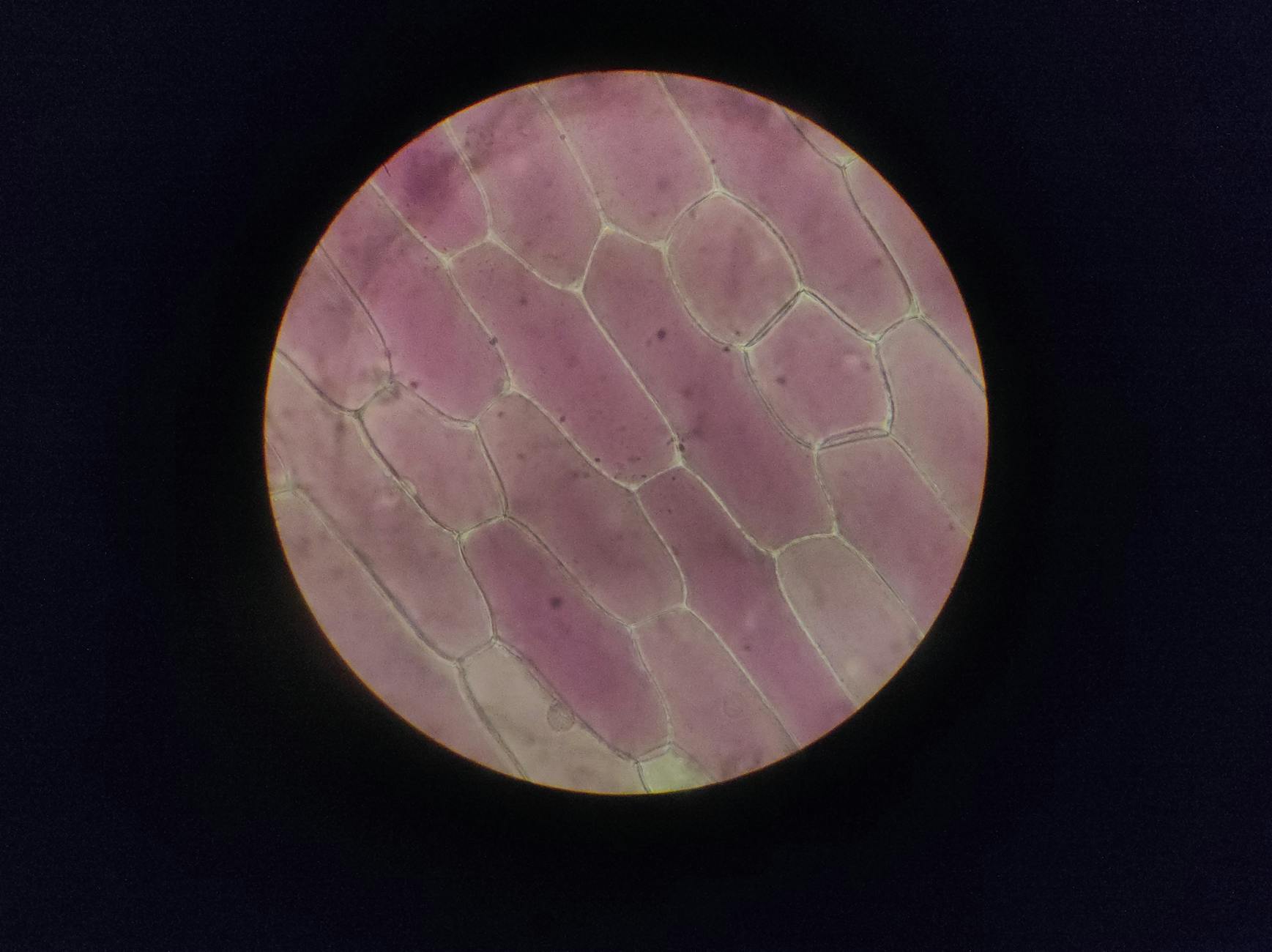
When a biopsy is taken from hypertrophic or keloid scar tissue, it shows microscopic signatures that explain why the scar appears raised and persistent. Pathologists observe thick bundles of hyalinized (glassy-looking) collagen arranged in characteristic whorls or nodular patterns. Crucially, biopsies reveal decreased collagenase activity—meaning the tissue is producing collagen faster than it can break it down. This imbalance is the core problem: the wound repair process doesn’t regulate itself properly, leading to excess collagen accumulation. Atrophic scars (the most common type after acne), which appear pitted or depressed, show a different pattern: loss of collagen and elastin in the dermis, creating the sunken appearance.
Biopsies of these scars reveal reduced collagen density compared to surrounding normal skin. Post-treatment biopsies are equally revealing. Studies show that 18 months after CO2 laser treatment, scar tissue shows increased collagen and elastin fibers, indicating that the treatment has stimulated the skin to actively remodel and repair the scarred area. This histological evidence is why lasers remain a first-line therapy—they trigger the body’s own healing mechanisms to regenerate healthy tissue. However, if scarring is very old or severe, collagen remodeling becomes slower and less reliable. In these cases, combination approaches (laser plus injectables, or surgical revision plus resurfacing) often yield better results than single treatments alone.
How Do Scar Types Correlate with Microscopic Findings?
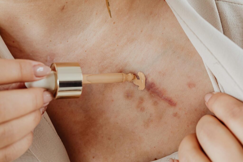
Dermatologists classify acne scars into distinct morphological types, and each has characteristic cellular features. Ice-pick scars are narrow, deep punctures with a concentrated loss of collagen. Rolling scars show broader depressions with sloped edges and tethering of deeper tissue to the skin surface. Box-car scars are wider, sharply demarcated depressions. These variations matter because they respond differently to treatment. A biopsy of an ice-pick scar would reveal a focal, deep collagen deficit, whereas rolling scars show broader tissue attachment changes that may require subcision (cutting the tethering beneath the scar) in addition to surface resurfacing.
Hypertrophic and keloidal scars present the opposite problem: overabundant collagen. A keloid biopsy distinctly shows collagen bundles extending beyond the original wound border, a key histological marker used to differentiate it from hypertrophic scarring. Biopsies also quantify fibroblast populations—the cells responsible for collagen production. Keloid tissue contains elevated numbers of fibroblasts with heightened collagen-synthesizing activity, explaining why keloids continue enlarging over time and why they are prone to recurrence even after treatment. Papular scars, raised but not deeply indented, show intermediate features: some collagen excess but without the organized whorled pattern of true hypertrophic scars. Understanding these distinctions guides treatment intensity and selection.
How Do Biopsy Findings Guide Treatment Selection?
The cellular architecture revealed by biopsy directly informs which treatment will be most effective. For hypertrophic and keloidal scars with excess collagen and reduced collagenase, laser therapy is highly effective because it vaporizes excess collagen and stimulates remodeling. Clinical evidence supports CO2 (10,600 nm wavelength) and Er:YAG (2,940 nm wavelength) lasers as first-line therapies, achieving up to 90% efficacy in reducing scar appearance. The mechanism is selective photothermolysis: the laser targets water in the tissue, creating controlled thermal injury that triggers the body to break down abnormal collagen and regenerate normal tissue. For atrophic scars with collagen loss, the treatment strategy differs.
Lasers still work, but the goal is different: fractional or ablative lasers resurface the skin and remodel remaining collagen while stimulating new collagen production via wound healing. Chemical peels, microneedling, and dermal fillers also address collagen deficits by either triggering new collagen synthesis or physically filling depressed areas. Biopsies demonstrating severely depleted collagen in atrophic scars support the use of regenerative approaches—injectables containing collagen-stimulating agents or emerging cell-based therapies that literally introduce new fibroblasts to repair the deficit. The tradeoff: aggressive treatments (ablative lasers, surgical excision) work faster but require longer recovery and carry higher risks of complications like persistent redness or pigmentation changes. Gentler approaches (microneedling, gentle peels, light lasers) require more sessions but suit sensitive skin and patients who cannot tolerate downtime.
What Advanced Biopsy Findings Inform Emerging Therapies?
Recent biopsy research has identified specific collagen and elastin patterns that guide next-generation scar treatments emerging in 2025–2026. Autologous cell-based therapies, such as those using stromal vascular fraction (SVF) derived from the patient’s own fat tissue, show promise because biopsies confirm that introducing living fibroblasts can regenerate organized collagen architecture rather than simply filling or resurfacing a scar. Similarly, ReCell® technology, which harvests a small skin biopsy from the patient and uses it to seed growth across scarred areas, leverages the body’s own tissue-repair capacity. Histological studies confirm that these approaches produce more durable, naturally-textured results compared to traditional scar treatments.
Topical biologics represent another frontier informed by biopsy science. Products containing peptides and growth factors target fibroblast dysfunction at the molecular level, supporting organized collagen production. These work best in conjunction with other treatments and typically require consistent use over months to show histological changes. However, for patients with widespread or severe scarring, or those unable to tolerate invasive procedures, emerging topical and injectable biologics offer an alternative path forward. A limitation is that topical products have limited skin penetration; they primarily affect the epidermis and superficial dermis, making them less effective for deep atrophic scars.

The Role of Scar Severity Grading in Treatment Planning
Dermatologists use a standardized 4-grade severity scale that correlates with biopsy findings and treatment expectations. Macular scars are red or flat but even with surrounding skin—these represent early inflammation with minimal collagen changes and often improve spontaneously or with light laser treatments. Mild scars are visible only with close examination and typically covered by makeup; biopsies show modest collagen abnormalities. Moderate scars are obvious at conversational distance and represent significant collagen loss or excess; these require multiple treatment sessions.
Severe scars are evident from more than 50 centimeters away and represent extensive tissue remodeling; these often demand combination approaches and may involve surgical revision. This classification system bridges clinical appearance with underlying histology. A dermatologist assessing your scars will place them within this framework to set realistic expectations. A patient with moderate atrophic scarring on the cheeks might need 4–6 laser sessions plus chemical peels and filler injections, whereas someone with mild rolling scars might achieve satisfactory improvement with 2–3 fractional laser treatments. Biopsy-informed understanding of collagen structure ensures treatment plans match the actual tissue pathology rather than relying on appearance alone.
Future Perspectives: How Biopsy Science Shapes Tomorrow’s Scar Treatments
As dermatology advances, biopsy research continues to reveal how to engineer better scarring outcomes at the cellular level. Regenerative medicine—leveraging the patient’s own cells to reconstruct damaged tissue—is moving from research to clinical practice. Biopsies demonstrating that cell-based therapies can reestablish normal collagen organization and elastin networks have validated this approach.
By 2026, we’re seeing expansion of injectables specifically designed for long-term collagen repair, drawing directly from histological evidence about what fibroblasts need to produce healthy, organized tissue rather than pathological scar collagen. The broader implication: future scar treatment may become increasingly personalized. Rather than applying a standardized protocol to all patients, dermatologists may eventually use biopsy data (or advanced imaging that mimics biopsy findings) to prescribe treatment tailored to each individual’s specific collagen architecture and fibroblast activity. This precision-medicine approach, grounded in the cellular insights that biopsies provide, promises to move beyond the current 60–90% efficacy rates toward more predictable, complete scar resolution.
Conclusion
A biopsy of acne scar tissue is not a standard diagnostic tool—clinical examination suffices for most patients. However, when performed, biopsies reveal whether scarring results from collagen excess (hypertrophic, keloidal) or collagen deficit (atrophic), fibroblast hyperactivity, and specific structural disorganization. These findings explain why certain treatments work better than others: lasers remodel excess collagen and stimulate regeneration, fillers and grafts address collagen loss, and emerging cell-based therapies literally introduce healthy fibroblasts to reconstruct organized tissue architecture. Understanding what biopsies reveal—while recognizing that your dermatologist can clinically diagnose and treat scars without one—empowers you to have informed conversations about your treatment options.
If you’re considering acne scar treatment, work with a board-certified dermatologist who can assess your scar type, severity, and skin characteristics to design a personalized plan. Laser therapy, chemical peels, microneedling, injectables, and surgical approaches all have robust clinical support. The newest regenerative therapies show genuine promise, particularly for severe or extensive scarring. Most patients benefit from combination treatments over time rather than a single procedure, and realistic expectations—understanding that scarring improves rather than disappears completely—support satisfaction with results.