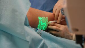
Wood’s lamp is used for fungal acne diagnosis because it reveals a distinctive fluorescence pattern that clearly distinguishes Malassezia folliculitis from bacterial acne. When exposed to the long-wave ultraviolet light emitted by a Wood’s lamp, Malassezia species produce a characteristic bluish-white or yellow-green fluorescence that is completely invisible to the naked eye. This fluorescence occurs because Malassezia produces pityrialactone, a tryptophan derivative metabolite that causes the organism to glow under UV light.
In contrast, bacterial acne caused by Cutibacterium appears orange-red under the same lamp, making differentiation between these two distinct conditions immediate and straightforward. The beauty of Wood’s lamp examination lies in its simplicity and reliability. A dermatologist can perform the test in seconds without any patient preparation, special equipment beyond the lamp itself, or waiting for culture results. This article explores why Wood’s lamp remains a standard diagnostic tool for fungal acne, how the technology works, the specific patterns that indicate infection, and what happens when a diagnosis is confirmed.
Table of Contents
- How Does Wood’s Lamp Reveal Fungal Acne?
- Clinical Advantages of Wood’s Lamp for Fungal Acne Diagnosis
- How Fluorescence Patterns Pinpoint Infection Sites
- Rapid Diagnosis Without Waiting for Culture Results
- Specimen Collection Precision and Diagnostic Yield
- Limitations of Wood’s Lamp Examination
- The Role of Wood’s Lamp in Modern Fungal Acne Management
- Conclusion
How Does Wood’s Lamp Reveal Fungal Acne?
Wood’s lamp uses long-wave ultraviolet light—specifically light at a wavelength of 365 nanometers—to illuminate fluorescent compounds that reflect visible light back to the observer’s eye. The human eye cannot see most of these fluorescent compounds under normal lighting, which is why the special lamp is necessary. When Malassezia colonizes hair follicles, the organism produces pityrialactone as a metabolic byproduct. This compound absorbs the ultraviolet light and re-emits it as visible light in the bluish-white to yellow-green spectrum, creating an unmistakable glow under Wood’s lamp examination.
The contrast between fungal and bacterial acne under Wood’s lamp is striking. Bacterial acne caused by Cutibacterium appears as an orange-red fluorescence due to the bacterial pigments present in the follicles, while Malassezia folliculitis glows blue-yellow. This visual distinction allows a clinician to differentiate the two conditions within moments of holding the lamp to the patient’s skin. The ability to see exactly which follicles harbor fungal infections also helps dermatologists pinpoint the precise locations for culture specimen collection, improving the accuracy of laboratory confirmation.
Clinical Advantages of Wood’s Lamp for Fungal Acne Diagnosis
Wood’s lamp examination is non-invasive and completely painless, requiring no needle sticks, swabs that might irritate skin, or patient preparation. The patient simply sits still while the dermatologist holds the lamp a few inches from the affected area and observes the fluorescence pattern. This ease of use makes it an ideal first-line diagnostic tool, particularly for patients who are uncomfortable with more invasive testing or who want rapid answers about their skin condition. Cost-effectiveness represents another major advantage.
Wood’s lamps are inexpensive instruments that have remained largely unchanged since their introduction decades ago, and dermatology offices routinely stock them. A Wood’s lamp examination costs significantly less than sending cultures to a laboratory, waiting for results, or performing genetic testing. However, one limitation to remember is that Wood’s lamp examination alone cannot identify which specific Malassezia species is causing the infection or rule out bacterial superinfection if both organisms are present. When species identification or mixed infections are suspected, culture remains necessary to complement the lamp findings.
How Fluorescence Patterns Pinpoint Infection Sites
The fluorescence patterns visible under Wood’s lamp provide a map of infection that guides specimen collection and treatment planning. When the lamp illuminates affected skin, the practitioner can see exactly which follicles glow with that characteristic bluish-white or yellow-green light. This visual clarity allows the clinician to collect culture specimens directly from actively infected sites rather than guessing where to swab, which dramatically increases the likelihood that culture samples will yield positive results for Malassezia.
For example, a patient presenting with scattered red bumps on the chest might appear to have typical acne to the naked eye, but Wood’s lamp examination might reveal that only certain follicles produce fluorescence. Those fluorescing follicles are the fungal-infected ones, while the others may be bacterial or inflammatory acne. This distinction is not merely academic—it changes treatment entirely. A patient with fungal acne treated with antibiotics will likely see little improvement and may develop resistance, whereas the same patient treated with antifungal medications will typically respond well.

Rapid Diagnosis Without Waiting for Culture Results
Traditional bacterial acne diagnosis often relies on clinical observation alone, with culture reserved for treatment-resistant cases. Fungal acne diagnosis, by contrast, benefits tremendously from the speed of Wood’s lamp examination. A patient can walk into a dermatology office, receive a diagnosis within minutes, and leave with a treatment plan—all without waiting three to seven days for culture results. This immediate feedback loop helps patients start appropriate antifungal therapy sooner, potentially shortening the duration of their infection.
The tradeoff, however, is that while Wood’s lamp provides rapid presumptive diagnosis, it cannot provide the detailed microbiological information that culture offers. If a clinician needs to know whether the patient harbors Malassezia globosa, Malassezia furfur, or another species, or if other organisms are also involved, culture is still necessary. Many dermatologists use Wood’s lamp as a screening tool and then order culture for confirmation or when treatment is not working as expected. This combined approach balances the speed of lamp examination with the definitive information that laboratory testing provides.
Specimen Collection Precision and Diagnostic Yield
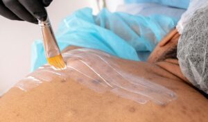
One practical advantage of Wood’s lamp that is often overlooked is its role in improving specimen collection accuracy. When a dermatologist uses the lamp to identify exactly which follicles are infected, they can target their culture swab or brush directly to those fluorescing areas. This targeted collection dramatically increases the diagnostic yield—meaning the culture is much more likely to grow Malassezia and provide definitive identification.
A patient with sparse, scattered fungal folliculitis might have numerous normal follicles interspersed among infected ones. Collecting a specimen from a non-infected follicle will yield negative culture results, potentially leading to diagnostic confusion. Under Wood’s lamp, the practitioner collects only from the glowing follicles, ensuring the sample truly contains the organism in question. This precision is particularly valuable in cases where fungal acne is suspected but not confirmed, because it reduces the chance of false-negative cultures that might lead to missed or delayed treatment.
Limitations of Wood’s Lamp Examination
While Wood’s lamp is valuable, it has genuine limitations that practitioners must understand. The examination works best on Malassezia species that produce adequate pityrialactone and on skin that is not heavily pigmented—in darker skin tones, the fluorescence may be harder to visualize, though it is still often present. Additionally, if the patient has recently applied topical products like sunscreen, cosmetics, or medications, the residue can interfere with visualization by reducing the contrast between fluorescing and non-fluorescing areas.
Another limitation is that Wood’s lamp cannot differentiate fungal acne from other conditions that might produce similar clinical appearances, such as rosacea or bacterial folliculitis in its early stages. The lamp reveals what fluoresces, but clinical judgment is still required to interpret the findings in context of the patient’s symptoms, distribution of lesions, and response to prior treatments. A dermatologist must always correlate lamp findings with the clinical presentation rather than relying on the lamp as a standalone diagnostic tool.
The Role of Wood’s Lamp in Modern Fungal Acne Management
Despite the availability of newer diagnostic techniques, Wood’s lamp examination has remained a standard in dermatology practice because it combines affordability, speed, and reliability. The lamp will not disappear from dermatology offices anytime soon, and learning to recognize the distinctive fluorescence patterns of Malassezia is part of dermatologic training. For patients seeking a straightforward, non-invasive way to determine whether their acne is fungal in nature, Wood’s lamp examination remains the most practical first step.
Looking forward, as awareness of fungal acne grows among patients and primary care physicians, Wood’s lamp examination will likely become even more common as a screening tool. More patients are now asking their providers, “Could this be fungal acne?” and Wood’s lamp provides an immediate, affordable answer. Combined with appropriate follow-up culture when needed and targeted antifungal treatment, this simple examination tool continues to play a vital role in improving outcomes for patients with Malassezia folliculitis.
Conclusion
Wood’s lamp serves as an essential diagnostic tool for fungal acne because it reveals the bluish-white to yellow-green fluorescence of Malassezia species through the production of pityrialactone, allowing immediate differentiation from bacterial acne. The examination is non-invasive, painless, rapid, cost-effective, and crucially, it pinpoints the exact location of fungal infections, which improves both diagnostic accuracy and specimen collection precision.
These advantages make it a reliable first-line tool that most dermatologists rely on when fungal acne is suspected. If you suspect you have fungal acne rather than bacterial acne—particularly if your breakouts have not responded to antibiotics or are concentrated in warm, moist areas like the chest or back—ask your dermatologist about a Wood’s lamp examination. This simple five-second test can confirm the diagnosis and guide your treatment toward appropriate antifungal medications rather than ineffective antibiotics, potentially saving you months of frustration and leading to faster healing.