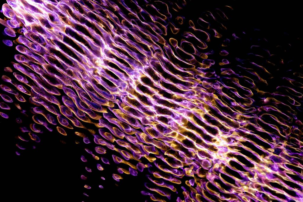
Reflectance confocal microscopy (RCM) reveals the microscopic architecture of acne-prone skin in unprecedented detail, showing inflammatory cell infiltration, bacterial colonization patterns, sebaceous gland involvement, and collagen disruption within the dermis and epidermis. In a patient with moderate acne, RCM can visualize individual immune cells surrounding a comedone, bacterial clusters within follicular structures, and areas of scar tissue formation that routine examination cannot detect. This article explores what dermatologists and researchers observe through RCM in acne skin, how these findings guide treatment decisions, and why this non-invasive imaging technology is becoming essential for understanding why some acne cases resist conventional therapy.
Table of Contents
- How Does Reflectance Confocal Microscopy Visualize Acne Pathology?
- What Inflammatory Changes Does RCM Reveal in Acne Skin?
- How Does RCM Visualize Bacterial Colonization in Acne Lesions?
- What Dermatologists Learn About Scarring Potential from RCM Findings?
- What Are the Limitations of RCM in Acne Assessment?
- How Does RCM Help Differentiate Acne from Similar Skin Conditions?
- The Future of RCM in Acne Management and Treatment Monitoring?
- Conclusion
- Frequently Asked Questions
How Does Reflectance Confocal Microscopy Visualize Acne Pathology?
RCM works by directing a focused laser beam into the skin and collecting the reflected light that bounces back from cellular and tissue structures. The technique achieves near-histological resolution—approximately 0.5 to 1 micrometer—without requiring a biopsy, making it uniquely suited for studying active acne lesions. As the laser scans horizontally across the skin and vertically through different depths, it creates real-time, optical cross-sections that reveal living cell arrangements, tissue organization, and pathological changes in precise detail. In acne lesions specifically, RCM shows a progression of changes from the epidermis downward.
At the follicular opening, the confocal image displays abnormally enlarged, hyperkeratotic epithelial cells that fill and occlude the pore. Moving deeper into the follicular wall, RCM reveals increased melanin content in keratinocytes and evidence of inflammation—bright, round immune cells infiltrating the tissue. Unlike traditional histology, RCM captures these cells in their living, active state rather than in the static, processed form of a biopsy specimen. This dynamic view shows whether immune cells are actively migrating into the lesion or already established within the follicular wall.
What Inflammatory Changes Does RCM Reveal in Acne Skin?
One of the most clinically valuable applications of RCM in acne is visualizing inflammatory cell populations without needing to excise tissue. In inflamed papules and pustules, RCM shows a characteristic “bright cell” pattern—these are primarily neutrophils and lymphocytes that appear as reflective cells against the darker background of normal dermis. In patients with severe cystic acne, RCM can differentiate between superficial inflammation (limited to the epidermis and upper dermis) and deep dermal inflammation (extending into the lower dermis), which has important implications for scarring risk and treatment selection.
However, RCM’s inflammatory findings don’t always correlate perfectly with clinical severity or treatment response. A patient might have extensive immune cell infiltration visible on RCM but respond rapidly to oral antibiotics, while another with apparently less dramatic confocal findings develops treatment-resistant disease. This discordance suggests that the quantity of inflammatory cells alone doesn’t determine acne severity—the type of cells present, their activation state, and their interaction with bacterial and lipid components matter significantly. Additionally, RCM’s depth limitation (it can only visualize approximately 350 micrometers into the skin) means very deep cystic lesions may not be fully characterized, potentially underestimating the inflammatory burden in severe nodular acne.
How Does RCM Visualize Bacterial Colonization in Acne Lesions?
Reflectance confocal microscopy can indirectly visualize bacterial populations within acne lesions by identifying areas of architectural disruption and inflammation characteristic of bacterial colonization. *Cutibacterium acnes* (formerly *Propionibacterium acnes*), the primary acne-associated bacterium, creates distinctive patterns within the follicular structure. RCM shows bacterial aggregates as regions of increased brightness and density within the follicle, often surrounded by inflammatory cells actively attempting to contain the infection.
A specific example is seen in patients with follicular impaction: RCM reveals tightly packed sebaceous material, colonizing bacteria, and inflammatory cells in a complex three-dimensional arrangement that explains why topical antibiotics alone often fail in these cases. The confocal images show that bacteria aren’t uniformly distributed throughout the lesion—they concentrate in specific microenvironments where sebum, follicular debris, and anaerobic conditions create protected niches. This visualization has driven research into combination therapies that both reduce bacterial load and disrupt the physical barriers that harbor bacteria, rather than relying solely on antimicrobial agents.
What Dermatologists Learn About Scarring Potential from RCM Findings?
RCM’s ability to assess collagen organization in the dermis has proven valuable for predicting scarring risk before clinically obvious scars develop. In normal skin, confocal images show organized, densely packed collagen bundles with a regular, parallel arrangement. In acne-affected skin, particularly after inflammatory lesions, RCM reveals disrupted collagen patterns, areas of collagen loss, and disorganized fibrous tissue that often precede visible atrophic or boxcar scarring.
The key advantage is timing: RCM can identify collagen disruption in the acute and subacute phases of acne healing, when intervention might prevent or minimize permanent scarring. A patient with nodular acne might show on RCM that deep dermal inflammation has already caused significant collagen disorganization, indicating that aggressive anti-inflammatory treatment and early consideration of resurfacing therapies would be appropriate. However, early RCM findings of collagen disruption don’t invariably predict clinical scarring—some patients with significant confocal changes heal without visible scars, likely due to individual variation in wound healing response and skin thickness. This means RCM should guide treatment decisions but not alone determine prognosis.
What Are the Limitations of RCM in Acne Assessment?
While RCM provides valuable information, several important limitations affect its clinical utility. First, the technique’s inability to visualize beyond approximately 350 micrometers depth means that deep nodular and cystic acne extending into the subcutis cannot be fully characterized. A severe cystic lesion might show significant inflammation in the upper and mid-dermis on RCM, but the full extent of the inflammatory process remains unseen. Second, RCM’s relatively slow acquisition speed compared to newer imaging modalities means that visualizing very extensive areas or multiple lesions becomes time-consuming, limiting its use as a rapid screening tool.
Additionally, operator experience significantly affects image quality and interpretation. A dermatologist inexperienced with confocal microscopy might acquire poor-quality images or misinterpret normal follicular structures as pathological findings, leading to unnecessary interventions. The learning curve for RCM is steeper than for clinical examination or standard photography. Furthermore, RCM works best on flat or minimally raised lesions and performs poorly on very inflamed, edematous, or pustular lesions where surface irregularity scatters light and degrades image quality. Some of the most clinically relevant acne lesions—active pustules and inflamed nodules—are precisely the lesions most difficult to image with RCM, which creates a practical constraint on when the technology can be meaningfully applied.

How Does RCM Help Differentiate Acne from Similar Skin Conditions?
RCM’s detailed visualization aids in distinguishing true acne from other folliculitis conditions and inflammatory disorders that may mimic acne clinically. Rosacea with follicular involvement, for example, shows characteristic RCM patterns of vascular dilation and a different distribution of inflammatory cells compared to acne lesions.
Fungal folliculitis from *Malassezia* or other organisms produces distinctive burrows and spore patterns visible on RCM that differ from bacterial acne. In a patient presenting with what appears to be widespread facial acne but resists conventional acne therapy, RCM can reveal alternative diagnoses—perhaps folliculitis from gram-negative bacteria resistant to standard acne antibiotics, or contact dermatitis producing acne-like lesions. This diagnostic clarity can redirect treatment entirely, from anti-acne regimens toward antibiotics targeting resistant organisms or elimination of a contact allergen.
The Future of RCM in Acne Management and Treatment Monitoring?
As RCM technology advances, faster acquisition speeds and improved imaging depth are expected to enhance its clinical utility in acne. Emerging three-dimensional confocal reconstruction techniques may provide better assessment of deep dermal involvement, improving scarring risk prediction.
Additionally, research is beginning to use RCM as an objective marker of treatment efficacy—measuring changes in inflammatory cell density, collagen reorganization, and bacterial distribution over weeks of therapy to determine whether a patient’s skin is responding at the cellular level, not just clinically. The potential for AI-assisted image analysis and automated pattern recognition in confocal microscopy images may eventually make RCM a more practical tool for widespread dermatologic use, moving it beyond specialized centers toward routine acne assessment. As this technology evolves, dermatologists may increasingly use RCM to guide early intervention in at-risk patients and to objectively monitor whether emerging treatments are actually improving the underlying pathology, not just surface appearance.
Conclusion
Reflectance confocal microscopy reveals that acne pathology extends far deeper and is far more complex than clinical examination can detect, showing inflammatory cell infiltration, bacterial colonization patterns, collagen disruption, and architectural changes within the follicle and dermis.
For dermatologists managing difficult-to-treat acne or attempting to assess scarring risk, RCM provides non-invasive, objective data that can guide treatment selection and predict outcomes. However, the technology’s limitations—depth restrictions, operator dependence, and performance challenges on very inflamed lesions—mean it functions best as a complementary tool alongside clinical examination rather than as a replacement for it.
Frequently Asked Questions
Can RCM be used to diagnose all types of acne?
No. RCM works best on non-pustular lesions and doesn’t effectively image very inflamed, edematous, or deeply situated nodular lesions. Clinical examination remains essential for comprehensive acne assessment.
Does RCM show individual bacteria in acne lesions?
RCM cannot visualize individual bacterial cells directly due to resolution limitations, but it shows areas of bacterial colonization as regions of increased brightness and density surrounded by inflammatory changes.
Can RCM predict whether acne will scar?
RCM can identify early collagen disruption and deep dermal inflammation that correlate with scarring risk, but individual healing responses vary, so RCM findings should guide treatment decisions but not serve as a definitive prognostic tool alone.
Is RCM available at all dermatology practices?
No. RCM remains a specialized tool available primarily at academic dermatology centers and some private practices. Equipment is expensive and requires operator training.
How long does RCM imaging take for a single lesion?
Typically 5 to 15 minutes per lesion depending on operator experience and lesion characteristics. Imaging multiple lesions can be time-consuming, limiting its use as a rapid screening tool.
Can RCM detect antibiotic-resistant bacteria in acne?
RCM cannot identify bacterial species or antibiotic resistance patterns directly. Culture or molecular testing is required for resistance assessment, though RCM can show that bacterial colonization persists despite antimicrobial therapy.