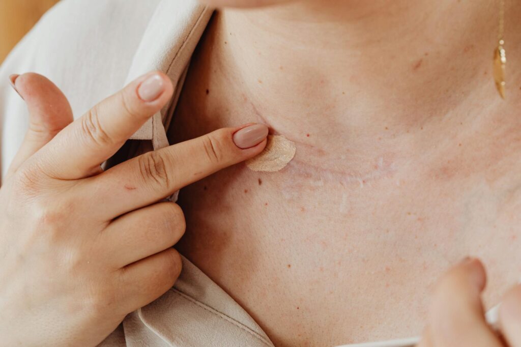
Acne scar consultations include photography and lighting assessment because scars behave differently under varying light conditions, and a dermatologist needs to see the full topographic picture before recommending treatment. A rolling scar that barely registers under soft overhead fluorescents can cast deep shadows under angled clinical lighting, revealing depth and texture that would otherwise go unnoticed. Without standardized photographic documentation taken under controlled lighting, a provider is essentially working from a single snapshot of a problem that changes appearance depending on the time of day, the room you are standing in, and even the angle of your head. Photography and lighting assessment together give the clinician a repeatable, objective baseline that guides treatment selection, tracks progress over subsequent visits, and protects both patient and provider by creating a visual medical record.
This process is not cosmetic vanity or a billing add-on. It is a diagnostic step rooted in the physical reality that atrophic and hypertrophic scars interact with light in measurably different ways. A boxcar scar, for instance, has sharply defined vertical edges that produce harsh shadow lines under cross-polarized lighting, while an ice pick scar may appear as little more than an enlarged pore under diffused light but reveals its true narrow depth under tangential illumination. The consultation photography session captures these distinctions so the clinician can classify each scar accurately and match it to the intervention most likely to improve it. This article covers how lighting conditions change scar visibility, why standardized clinical photography matters for treatment planning, what specific lighting techniques practitioners use, how to prepare for this part of your consultation, and the limitations patients should understand about photographic assessment.
Table of Contents
- How Does Lighting Change the Way Acne Scars Appear During Consultation?
- Why Standardized Clinical Photography Is Essential for Accurate Scar Classification
- Specific Lighting Techniques Used in Acne Scar Assessment
- How to Prepare for the Photography Portion of Your Scar Consultation
- Limitations and Common Misunderstandings About Photographic Scar Assessment
- How Photography Documentation Supports Long-Term Treatment Tracking
- Where Scar Assessment Technology Is Heading
- Conclusion
- Frequently Asked Questions
How Does Lighting Change the Way Acne Scars Appear During Consultation?
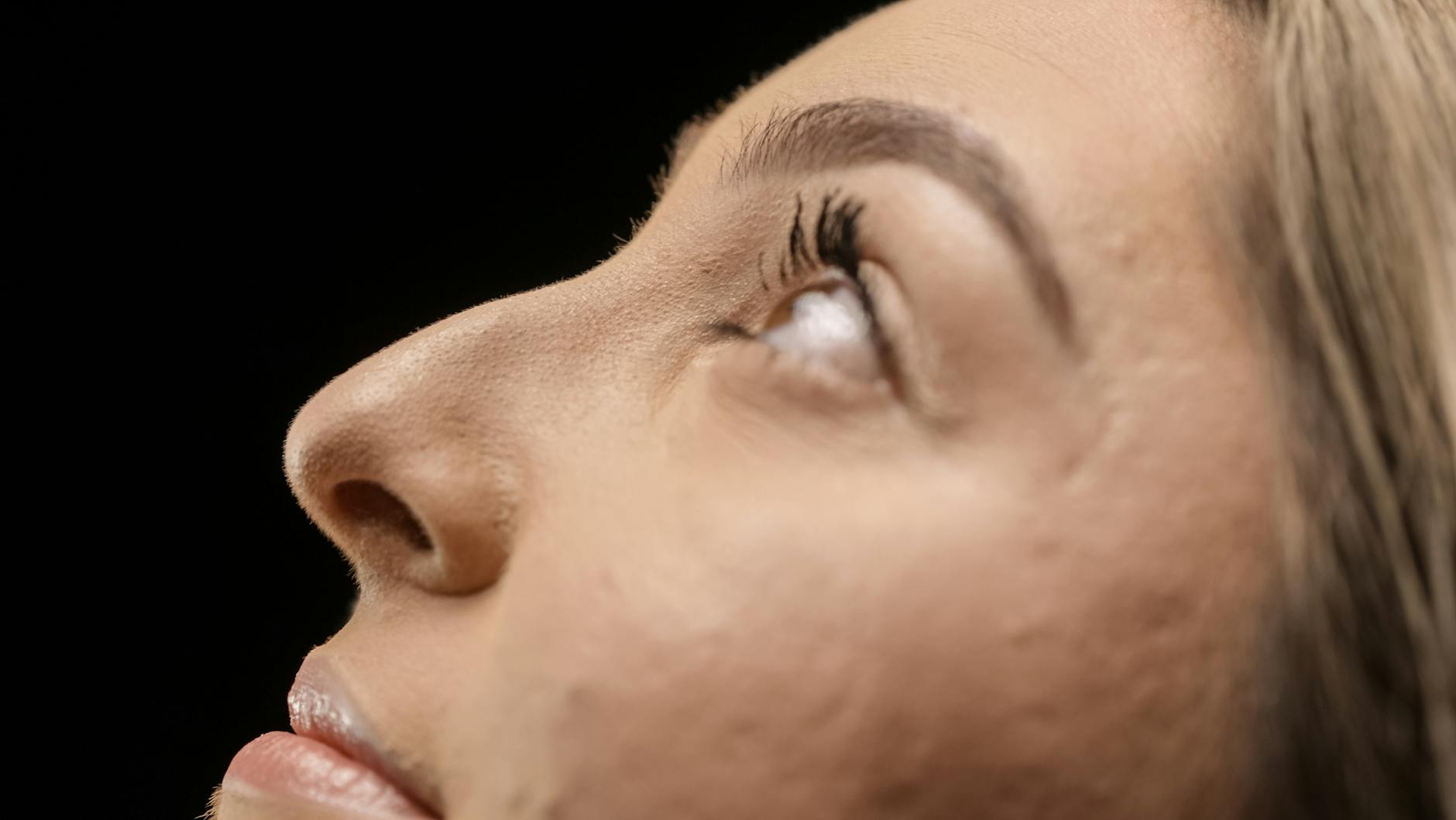
Light does not simply illuminate acne scars — it defines them. The visibility of a scar depends almost entirely on the angle, intensity, and quality of the light hitting the skin surface. Dermatologists have understood this since at least the 1980s when standardized clinical photography protocols began appearing in plastic surgery literature, but many patients are surprised to learn how dramatically a scar’s apparent severity shifts between lighting conditions. Under direct frontal flash, which is what most bathroom mirrors and smartphone selfies approximate, shallow rolling scars can virtually disappear because the light fills in the depressions evenly. Move that same patient under raking light positioned at a fifteen to thirty degree angle from the skin surface, and those rolling scars suddenly throw visible shadows that reveal their true width and depth. This is not an illusion in either direction — it is the same scar seen two different ways, and the clinician needs both views. The comparison matters because treatment decisions hinge on scar morphology.
A 2019 classification study published in the Journal of Clinical and Aesthetic Dermatology reinforced that scar depth, edge sharpness, and surface texture are the three variables most predictive of treatment response, and all three are lighting-dependent observations. A provider who only examines your skin under the flat fluorescent panels typical of a general practice exam room may underestimate scar severity and recommend a superficial treatment like a light chemical peel when subcision or fractional laser resurfacing would be more appropriate. Conversely, a consultation room with only harsh angled lighting might overstate the severity of shallow texture irregularities, leading to unnecessarily aggressive treatment. The photography and lighting assessment exists to prevent both errors by capturing the scar field under multiple controlled conditions. Cross-polarized photography adds another diagnostic layer that the naked eye cannot replicate. Standard photography captures surface reflections along with subsurface detail, which can obscure vascular components of scarring — the persistent redness or hyperpigmentation that often accompanies acne scars. Cross-polarized filters eliminate surface glare and reveal the chromatic component of scarring independently from the textural component. This distinction matters because a scar that is both depressed and red may need a combination approach — resurfacing for the depression and vascular laser or topical treatment for the discoloration — and separating those two features visually during the consultation helps the provider build a layered treatment plan rather than addressing only what is most obvious under a single lighting condition.
Why Standardized Clinical Photography Is Essential for Accurate Scar Classification
Dermatologists classify acne scars using systems like the Goodman and Baron qualitative scale or the more granular quantitative ECCA grading system, and both rely on visual assessment that is only as reliable as the viewing conditions. Standardized photography means consistent camera distance, fixed lighting positions, identical exposure settings, and reproducible patient positioning across every session. Without this standardization, comparing a baseline photo to a three-month post-treatment photo becomes unreliable, because differences in lighting angle or camera distance can make scars look better or worse independently of any actual tissue change. A patient who tilts their chin slightly higher in the follow-up photo may appear improved simply because the overhead light no longer catches the scar margins the same way. However, standardization has a real limitation that patients should understand: even the most rigorous photographic protocol captures a two-dimensional representation of a three-dimensional problem. Photography compresses depth information, which is why some clinics supplement standard photography with three-dimensional imaging systems like Canfield VECTRA or Antera 3D.
These systems use structured light projection to generate topographic maps of the skin surface, measuring scar depth in micrometers rather than relying on shadow interpretation. If your consultation includes only two-dimensional photography, the lighting assessment becomes even more critical because the clinician is depending on shadow behavior to infer depth — a task that requires deliberate, multi-angle illumination rather than a single well-lit photograph. The documentation value also extends beyond treatment planning. Standardized before-and-after photography protects patients by creating an objective record of baseline severity, which matters if a treatment causes an unexpected adverse reaction or if results fall short of what was discussed. It equally protects the provider from disputes about whether improvement occurred. clinics that skip this step or take only casual smartphone photos under variable room lighting are cutting a corner that can create problems months down the line when memory of the original scar severity has faded and the only evidence is a poorly lit snapshot taken at an inconsistent angle.
Specific Lighting Techniques Used in Acne Scar Assessment
The most common clinical lighting technique for scar evaluation is tangential or raking light, where the light source is positioned nearly parallel to the skin surface. At angles between ten and thirty degrees, this technique maximizes shadow formation in depressed scars and highlights raised edges in hypertrophic or keloidal scars. Practitioners typically use a moveable arm-mounted LED panel that can be repositioned during the examination. A dermatologist evaluating a patient with mixed scarring across the cheeks might start with the light source at the patient’s ear level, sweeping it slowly forward while observing how shadows form and shift across the scar field. This dynamic assessment often reveals scars that static overhead lighting misses entirely — particularly shallow rolling scars that may cover a broad area but have gentle slopes that only cast visible shadows at specific angles. Diffused frontal lighting serves as the complementary baseline. This is the even, shadow-minimizing illumination that shows the skin as most people see it in everyday conditions. Clinically, it is useful for assessing pigmentary changes — post-inflammatory hyperpigmentation and erythema — because color variations are easier to evaluate without competing shadow artifacts.
Some clinics use ring lights or dual softbox setups to achieve this uniform illumination. The comparison between what a scar looks like under diffused light versus raking light is itself diagnostic information. A scar that looks severe under raking light but nearly invisible under diffused light is primarily a textural issue, while a scar that remains prominent under both conditions likely has significant pigmentary involvement alongside the structural depression. Ultraviolet fluorescence photography, specifically Wood’s lamp examination, occasionally appears in acne scar consultations, though it is more commonly associated with active acne assessment. Under UV illumination, certain skin characteristics fluoresce differently, which can reveal the extent of subsurface inflammation or residual bacterial activity in the scar bed. This technique is most useful when the provider suspects that what appears to be static scarring may still have an active inflammatory component, which would change the treatment timeline. Treating inflammatory lesions with aggressive resurfacing can worsen outcomes, so identifying active inflammation under UV light can prevent a premature procedural intervention. That said, not every clinic uses UV photography for scar consultations specifically, and its absence does not indicate a substandard evaluation.

How to Prepare for the Photography Portion of Your Scar Consultation
Arrive at your consultation with clean, bare skin — no makeup, tinted moisturizer, sunscreen with a white cast, or concealer. This seems obvious, but it is the single most common issue that compromises consultation photography. Even a thin layer of silicone-based primer can partially fill shallow scars and alter the way light interacts with the skin surface. Some clinics will ask you to wash your face on site, but arriving bare-faced ensures the photography reflects your actual skin texture without any product interference. If you wear makeup to work and have an afternoon appointment, bring makeup remover and cleanse thoroughly before the session rather than relying on a quick splash of water in the clinic restroom. The tradeoff patients should consider is between convenience and accuracy. A consultation scheduled during your lunch break may mean rushing through cleansing, arriving with residual product on the skin, or having post-cleansing redness from aggressive makeup removal that the camera then captures as part of your baseline.
Scheduling the consultation as a first appointment of the day, when your skin has only had morning cleansing and no product application, produces the most representative photographs. Similarly, if you have been using a retinoid or exfoliating acid that causes visible peeling, the flaking skin can create its own shadow artifacts that interfere with scar assessment. Some providers recommend pausing harsh actives for three to five days before a photographic assessment, though this varies by practice and should be confirmed when booking. Bring your own photos if you have them — especially photos taken in different lighting conditions or at different times of day. These are not a substitute for clinical photography, but they provide useful supplementary information. A patient who says “my scars look worst in late afternoon car lighting” is identifying a specific lighting angle and color temperature that bothers them most, and that subjective concern informs the clinical assessment. Your goals matter alongside the objective findings. A scar that is clinically shallow but catches afternoon sunlight in a way that makes you self-conscious at work is still a valid treatment target, and your casual photographs help the provider understand which scars create the most distress in your actual daily lighting environments.
Limitations and Common Misunderstandings About Photographic Scar Assessment
The most important limitation of clinical photography, even under controlled lighting, is that photographs cannot fully capture how scarring looks in motion. Skin moves, stretches, and compresses throughout the day as you talk, smile, and turn your head. A scar that appears minimal in a static photograph may become conspicuous when the surrounding skin moves because the scar tissue does not flex the same way as unscarred skin. This is particularly relevant for tethered scars — depressed scars that are anchored to deeper tissue layers and pull downward during facial movement. Subcision is often the recommended treatment for tethered scars, but the tethering may not be apparent in any static photograph regardless of lighting. Providers who assess scars only from photographs without also observing the patient’s skin in motion may miss this component. Another misunderstanding is that better photography equipment automatically means a more accurate assessment.
While higher resolution cameras and specialized lighting systems produce more detailed images, the clinical interpretation still depends on the provider’s training and experience. A dermatologist with fifteen years of scar revision experience using a basic DSLR and a single adjustable light source will generally produce a more accurate assessment than a med spa using an expensive 3D imaging system operated by a technician without deep scar morphology training. The technology supports the assessment — it does not replace clinical judgment. Patients should be cautious about clinics that lean heavily on impressive imaging technology in their marketing without clearly explaining how those images translate into specific treatment decisions. The question to ask is not “what camera system do you use” but “how does the photography change what you recommend.” Skin tone also introduces a variable that lighting alone cannot fully resolve. On deeper skin tones, shadow-based scar assessment can be less reliable because the contrast between shadowed and illuminated skin is lower, and post-inflammatory hyperpigmentation can visually mask or exaggerate textural irregularities depending on the lighting angle. Experienced providers adjust their lighting protocols for different skin tones, often relying more heavily on cross-polarized and tangential techniques in combination rather than any single method. If you have a darker skin tone and your consultation feels rushed or uses only one lighting setup, it is reasonable to ask whether additional imaging could improve the assessment accuracy.

How Photography Documentation Supports Long-Term Treatment Tracking
Scar treatment is rarely a single-session event. Most patients undergo a series of treatments — often combining modalities like microneedling, fractional laser, subcision, and filler — spaced weeks or months apart. Standardized photography at each visit creates a longitudinal visual record that allows the provider to measure incremental improvement that may not be obvious visit to visit.
A patient four sessions into a fractional CO2 laser series may feel discouraged because they still see scars in the mirror every morning, but side-by-side comparison of their baseline photographs against the current session’s images, taken under identical lighting conditions, often reveals meaningful improvement in scar depth and shadow behavior that daily self-observation cannot detect. This documentation also informs mid-course treatment adjustments. If photographs show that rolling scars are responding well to fractional resurfacing but ice pick scars remain largely unchanged after three sessions, the provider has visual evidence to justify pivoting the treatment plan — perhaps adding TCA CROSS for the ice pick component while continuing resurfacing for the broader texture. Without consistent photographic documentation, these nuanced comparisons rely on memory and subjective impression, which are unreliable over treatment courses that may span six to twelve months.
Where Scar Assessment Technology Is Heading
The integration of artificial intelligence into clinical photography is beginning to change how scar assessment works at the consultation stage. Several research groups and commercial platforms are developing machine learning models trained on large datasets of scar photographs to assist with automated classification and severity grading. These systems analyze shadow patterns, texture metrics, and color data from standardized photographs and return a quantitative scar map that supplements the clinician’s visual assessment. Early validation studies have shown promising agreement between AI-generated scar grades and expert consensus ratings, particularly for distinguishing between scar subtypes — boxcar versus rolling versus ice pick — which is one of the more subjective elements of manual classification.
The practical implication for patients in the near term is that consultation photography protocols may become more standardized across clinics as AI-assisted analysis requires consistent input data to produce reliable output. This could reduce the current variability in consultation quality between providers who invest in rigorous photography workflows and those who do not. However, these tools remain supplementary, not diagnostic, and the regulatory landscape around AI-assisted dermatologic assessment is still developing. For now, the best consultation is still one where a knowledgeable provider uses thoughtful lighting and photography to see your scars as completely as possible, documents what they find, and explains clearly how those findings translate into a treatment plan built around your specific scar morphology and goals.
Conclusion
Photography and lighting assessment during an acne scar consultation is a clinical necessity, not an upsell or a formality. The interaction between light and scarred skin is complex enough that a single viewing condition — whether it is your bathroom mirror, your phone’s front camera, or a clinic exam room’s overhead panel — will always give an incomplete picture. Tangential lighting reveals depth. Cross-polarized imaging separates color from texture.
Standardized photography creates a reliable baseline for tracking treatment progress. Together, these techniques give your provider the visual information needed to classify your scars accurately, select appropriate treatments, and measure whether those treatments are working over time. If you are scheduling an acne scar consultation, look for a provider who takes the photography component seriously — dedicated lighting equipment, consistent positioning protocols, and the willingness to explain what the images reveal about your specific scarring. Arrive with clean bare skin, bring any of your own photos that show your scars under conditions that bother you most, and ask how the photographic findings will influence the treatment plan. The lighting assessment is where a competent scar consultation separates itself from a superficial one, and it is worth understanding what that process involves before you sit down in the chair.
Frequently Asked Questions
Does insurance cover the photography and lighting assessment portion of an acne scar consultation?
In most cases, acne scar treatment is classified as cosmetic, and the consultation photography is bundled into the consultation fee rather than billed separately. Some exceptions exist when scarring results from a documented medical condition or injury, but patients should assume the photography cost is part of the out-of-pocket consultation fee and confirm with the clinic before booking.
How long does the photography and lighting assessment portion of the consultation typically take?
The photography itself usually takes ten to twenty minutes, depending on the extent of scarring and how many facial zones need documentation. The full consultation, including the visual assessment discussion and treatment planning, typically runs thirty to sixty minutes total. Clinics using 3D imaging systems may take slightly longer due to calibration and image processing time.
Can I take my own photos at home under different lighting to bring to my consultation?
Yes, and many providers encourage it. Photos taken in natural side-lighting near a window, under overhead bathroom lighting, and in outdoor daylight each reveal different scar characteristics. These will not replace clinical photography but they show the provider which conditions make your scars most visible to you, which helps calibrate treatment goals to your real-world experience.
Will my consultation photographs be shared or used for marketing purposes?
Not without your explicit written consent. Medical photographs are part of your health record and are subject to privacy protections. Some clinics may ask permission to use anonymized before-and-after images for educational or marketing purposes, but you are never obligated to agree, and declining will not affect your care.
Why do my scars look worse in the clinical photos than they do to me at home?
Clinical lighting is specifically designed to maximize scar visibility for diagnostic accuracy. The raking light and cross-polarized techniques used in consultations reveal detail that everyday lighting conditions typically soften or hide. The clinical photos show the maximum extent of scarring — your daily experience under normal lighting is also real, and treatment goals are usually set with your everyday appearance in mind, not the worst-case clinical view.